
Narrative Review · Bone Tissue Engineering
 Bioengineering (MDPI)
Scaffolds Built for
Osteoporotic Bone.
Bone defects in osteoporosis are still repaired with grafts and scaffolds validated in healthy bone. This review asks what a 3D-printed or biofabricated scaffold would need to do differently — and how strong the evidence is for each answer.
4
Design Axes
5.5%
Subsidence, Porous Ti
62
References
Why This Review Was Needed
Not rarefied bone — a different environment.
Fragility fracture is a growing global burden. Hip fractures alone are projected to nearly double in absolute number between 2018 and 2050, with a steeper rise in men (2.4-fold) than women (1.7-fold). Yet autograft is limited by donor-site morbidity and depends on host biology that osteoporosis compromises, and solid metal implants are far stiffer than the bone they sit in.
Earlier reviews treated materials, immune regulation and the redox–senescence axis separately. To the authors’ knowledge, none brought mechanical adaptation, redox regulation, senescence modulation and immunovascular coupling into one design framework for osteoporotic bone — or systematically separated tested evidence from design hypothesis.
172.79M
New fractures worldwide in 2021
453.31M
Prevalent fracture cases
25.18M
Years lived with disability
Global Burden of Disease Study 2021.
How osteoporotic bone differs
Cortical thinning and trabecular loss
Poor host bone for fixation and impaired load transfer.
Adipogenic progenitor bias
Ageing and oestrogen withdrawal push marrow stromal cells towards fat rather than bone, so scaffolds relying on endogenous progenitors draw on a diminished reservoir.
Senescent cells and their secretory phenotype
An anti-regenerative, pro-inflammatory local milieu — interleukin-6, interleukin-1, TNF-α, matrix metalloproteinases and chemokines.
Type-H vascular decline
Loss of the CD31-high/endomucin-high capillaries that couple angiogenesis to osteogenesis.
Inflammageing and immunosenescence
Persistent inflammation and impaired resolution.
Blunted mechanotransduction
In aged bone the same mechanical stimulus may produce less new bone — the effective anabolic threshold appears to rise.
Four Design Axes
Four jobs for one construct.
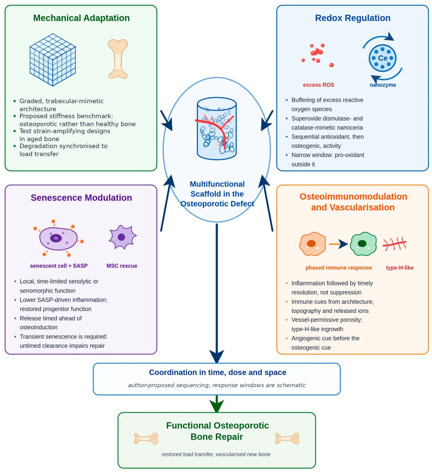
Additive manufacturing and biofabrication can do what passive grafts cannot: patient-specific geometry from CT data, internal architecture graded to mimic trabecular bone, and control over where and when biological cues are released. The review appraises printed scaffolds against four interdependent axes.
Mechanical adaptation
Graded, trabecular-mimetic architecture that restores stability in poor host bone without the stress shielding of solid high-modulus metal. The authors propose benchmarking stiffness to osteoporotic rather than healthy bone, and testing strain-amplifying designs.
Redox regulation
Local buffering of excess reactive oxygen species — for example with cerium oxide nanoparticles, whose surface chemistry mimics superoxide dismutase and catalase. The window is narrow: nanoceria can turn pro-oxidant in acidic environments.
Senescence modulation
Local, time-limited senolytic or senomorphic function to lower SASP-driven inflammation and restore progenitor function. Transient senescence is part of normal repair, so untimed clearance can impair healing.
Osteoimmunomodulation and vascularisation
Restoring the timing of inflammation and resolution rather than suppressing it, with vessel-permissive porosity and angiogenic cues ahead of the osteogenic cue.
The Proposed Triad
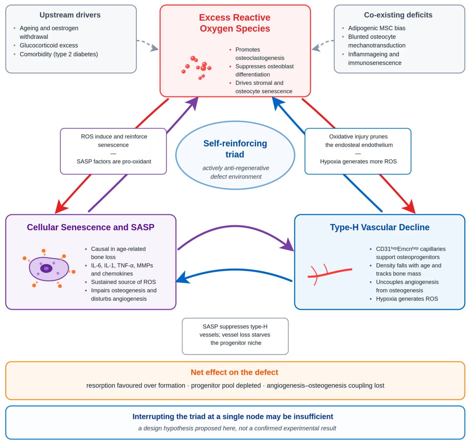
Three processes that feed each other.
Excess reactive oxygen species drive bone resorption and push stromal cells and osteocytes into senescence. Senescent cells, in turn, are a sustained source of oxidants — a self-amplifying redox–senescence loop already recognised in skeletal ageing.
The review adds a third node: the decline of type-H vessels, the capillaries that support bone-forming progenitors. Oxidative and senescent signalling suppress that endothelium, and hypoxia from vascular loss generates more oxidants.
If the three reinforce one another, a scaffold that interrupts only one of them may not be enough. The authors present this as a design hypothesis, not a confirmed mechanism.
Where the Evidence Stands
Mechanical signal is clinical.
The rest is preclinical.
The review labels every claim by study type and directness — whether it was tested in an osteoporotic model, a healthy one, or not at all. Within the searches described, the authors identified no completed clinical evaluation with reported results of a multifunctional scaffold in an osteoporosis-defined cohort.
| Axis | Most direct evidence | Setting and directness |
|---|---|---|
| Mechanical | Retrospective clinical cohort: 124 patients, 163 lumbar interbody fusion levels. Subsidence in 5.5% of porous printed titanium levels (4/73) versus 24.4% of solid titanium levels (22/90). | Clinical association only — patients were not selected for osteoporosis, and does not establish reduced stress shielding as the mechanism. |
| Redox | Cerium-bearing 3D-printed bioactive-glass and hydroxyapatite scaffolds gave early antioxidant effects followed by enhanced osteogenesis in rats. | Small-animal, non-osteoporotic. Untested in human osteoporotic bone; dose–response, biodistribution and clearance poorly defined. |
| Senescence | A sequentially releasing construct — dasatinib plus quercetin in GelMA with BMP-2 bioglass microspheres — improved regeneration of a 2.5 mm femoral defect in ovariectomised rats. | Small-animal, osteoporotic. Human evidence is for the systemic drug only, and its phase 2 trial missed its primary endpoint. |
| Osteoimmunomodulation | Porous 3D-printed Ti6Al4V releasing icariin and Mg²⁺ shifted macrophages towards M2 and improved peri-implant bone in osteoporotic rats. | A single small-animal osseointegration study — not defect repair, and not human evidence. |
The one human senolytic trial
In a phase 2 randomised trial of intermittent dasatinib plus quercetin in 60 postmenopausal women, the primary endpoint — 20-week change in the bone-resorption marker CTx — did not differ between groups (p = 0.611). The bone-formation marker P1NP rose 16% relative to control at two and four weeks, but not at 20. The authors read this transient signal as consistent with a time-dependent response, while noting a single regimen cannot establish a dose–response shape — and that it tested a systemic drug, not a scaffold.
A Time-Sequenced Framework
Timing, not more bioactivity.
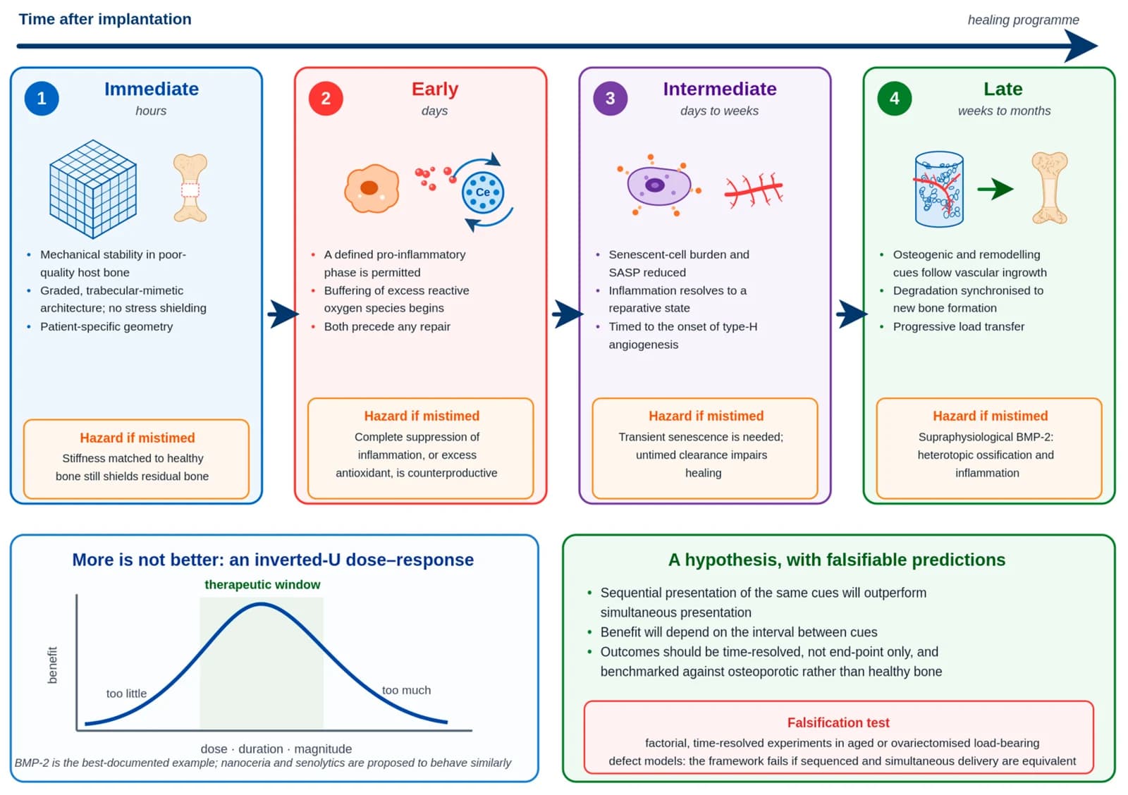
The central hypothesis: a scaffold for osteoporotic bone may depend less on how many biological functions it carries than on their coordination in time, dose and space. The authors propose engaging the axes sequentially rather than all at once.
Phase 01
Immediate · hours
Stabilise
Mechanical stability in poor-quality host bone from graded, patient-specific architecture, without stress shielding.
Hazard if mistimed
Stiffness matched to healthy bone may still shield residual bone.
Phase 02
Early · days
Buffer
Early redox buffering that accompanies — rather than abolishes — the necessary inflammatory phase.
Hazard if mistimed
Complete suppression of inflammation, or excess antioxidant, is counterproductive.
Phase 03
Intermediate · days to weeks
Resolve
As inflammation subsides, time-limited senescence and immune modulation paired with vascular support.
Hazard if mistimed
Transient senescence is needed; untimed clearance impairs healing.
Phase 04
Late · weeks to months
Build
Osteogenic and remodelling cues follow vascular ingrowth, with degradation synchronised to new bone for progressive load transfer.
Hazard if mistimed
Supraphysiological BMP-2 has caused heterotopic ossification and inflammation.
More is not better — the inverted U
Many interventions may work only inside a window of dose and time and do harm outside it. BMP-2 is the best-documented example: in a segmental defect model, supraphysiological doses induced heterotopic ossification and inflammation. The authors use the inverted U as a design heuristic, not a demonstrated osteoporotic dose–response.
How the framework could be proved wrong
It predicts that sequential presentation of the same cues will outperform simultaneous presentation, and that the benefit will depend on the interval between them. Factorial, time-resolved experiments in aged or ovariectomised load-bearing defect models would falsify it if sequenced and simultaneous delivery proved equivalent.
From Bench to Patient
The barriers are not only biological.
Bioink and batch consistency, sterilisation compatibility, cell sourcing and shelf life, tumorigenicity of cell-based components, long-term degradation and fatigue, and regulatory classification of combination and point-of-care products all stand between a promising construct and clinical use.
The review argues sterilisation should be fixed early as a design parameter, and that these constraints are as influential as any aspect of biological design.
A minimum validation package, and other priorities
- Osteoporosis-defined animal models with a healthy comparator and a clinically relevant, load-bearing defect
- Aged and comorbid — diabetic, glucocorticoid-exposed — large-animal models, to reduce reliance on young ovariectomised rodents
- Both sexes, or justified sex selection: most models study females, yet the steepest projected rise in hip fracture is in men
- Time-resolved biological endpoints alongside mechanical and fatigue testing, with follow-up long enough to assess degradation
- Early-phase trials with explicit osteoporotic selection and standardised outcomes
- Routine reporting of negative, null and dose–response results
What This Review Does Not Show
Promising, but clinically unproven.
The authors conclude that multifunctional scaffolds remain scientifically promising but clinically unproven for osteoporotic bone. The review produced no new data; it appraises published work and proposes a model to test.
A narrative review, not a systematic one
No protocol was registered, study selection was not duplicated, no formal risk-of-bias assessment or meta-analysis was performed, and no PRISMA-style screening log was kept. “Not identified” means not found within the searches described, not proof of absence.
A thin disease-specific evidence base
Osteoporosis-specific in vivo work is dominated by young ovariectomised rodents with small, non-load-bearing defects and short follow-up. Male subjects are largely absent, and much of the redox and immunomodulatory evidence comes from non-osteoporotic models.
Not all of it is additive manufacturing
Several of the most osteoporosis-specific constructs — strontium-doped bioceramics and cements, senolytic-releasing hydrogels — were made by conventional fabrication, so they inform scaffold design more directly than they validate 3D printing.
The framework is a hypothesis
The redox–senescence–vascular triad, the time-sequenced framework and the inverted-U argument are author-proposed syntheses. Individual elements are supported by the cited literature, but the framework as a whole is untested. The figures are schematic, not measured data.
Proposed, not prescribed.
A testable, time-sequenced design model for osteoporotic bone scaffolds — with every claim labelled by how directly it has been tested.
Review Identity
- Authors:
- Sedeek Mosaid · Yousif Jihad · Nuala Pepper · Mohamed Elnemr · Ashok Marudanayagam · Paul Lee
- Institution:
- Orthopaedic Surgery Department, United Lincolnshire Hospitals NHS Trust, Lincoln
- Journal:
- Bioengineering 13(9), published September 2026
- Article:
- 1088 · Review
- Method:
- Narrative review; PubMed/MEDLINE, Scopus, Web of Science and trial registries searched to August 2026
- Licence:
- Creative Commons Attribution (CC BY)
Citation
Mosaid S, Jihad Y, Pepper N, Elnemr M, Marudanayagam A, Lee P. 3D-Printed and Biofabricated Scaffolds for Osteoporotic Bone Defect Repair: Mechanical Adaptation, Redox Control, Senescence Modulation and Osteoimmunomodulation. Bioengineering. 2026;13(9):1088. doi:10.3390/bioengineering13091088
The authors declare that the research received no external funding and that they have no conflicts of interest. Figures reproduced from the paper under its CC BY licence.
Funding and affiliations
Affiliations: Orthopaedic Surgery Department, United Lincolnshire Hospitals NHS Trust
Funding: No external funding.
Publication: https://doi.org/10.3390/bioengineering13091088